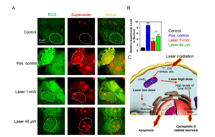
Fig. 7.
Low dose laser irradiation induces nuclear accumulation of superoxide. (A) Cells were labeled with the ROS-sensitive fluorescent dyes using the cellular ROS/Superoxide detection kit (Abcam, Cambridge, United Kingdom) and then treated by different laser fluences. The fluorescence images were acquired by confocal microscopy. Non-irradiated cells with no chemical treatment served as control. Pyocyanin (200 μM) treatment was used in the non-irradiated cells representing the ROS + positive control. (B) Quantitative analysis of relative superoxide fluorescence emission intensity from cells treated by laser. ImageJ software (NIH) was used for image processing and quantification. Data are expressed as means ± SEM (n = 3), **P< 0.01 ***P< 0.001. (C) Scheme of district biochemical signaling activation in cells after stimulation with different laser doses. ∆mΦ – mitochondrial membrane potential.

