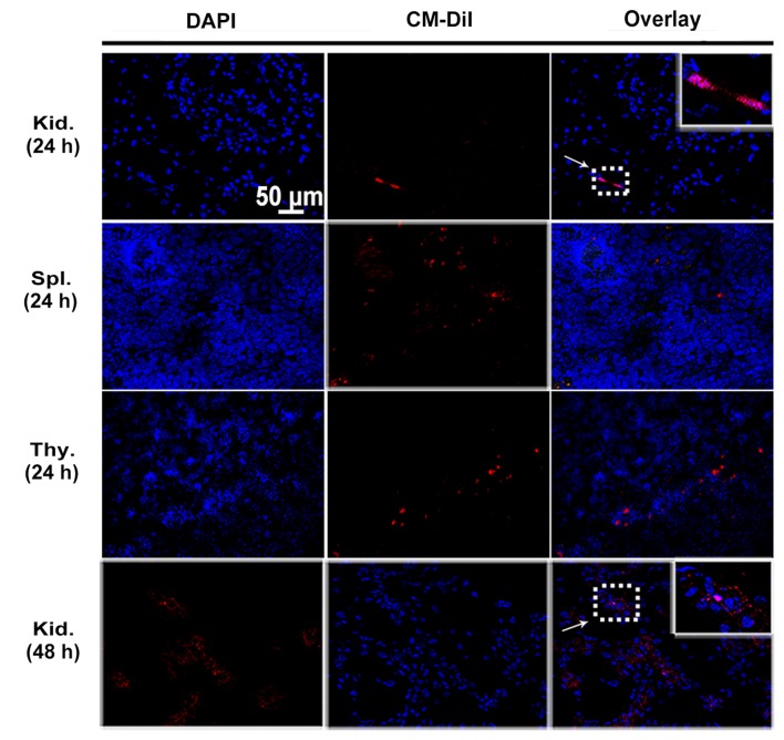
Figure 4.
In vivo tracking of engrafted mesenchymal stem cells (MSCs) at 24 and 48 h. Representative images of kidney (Kid.), spleen (Spl.) and thymus (Thy.) sections from diabetic rats injected with 5×10⁶ of CM-DiI labeled MSCs at 24 and 48 h after cell infusion. MSCs labeled with CM-DiI showed red fluorescence (arrow), and nuclei were stained by DAPI with blue fluorescence. Magnification, ×400.

