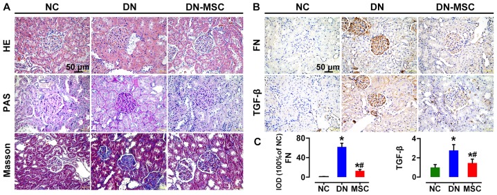
Figure 6.
Effect of mesenchymal stem cells (MSCs) on renal histopathological changes and expression of fibronectin (FN) and transforming growth factor-β (TGF-β) at 10 weeks after diabetes onset. (A) Representative images of hematoxylin and eosin (H&E), periodic acid-Schiff (PAS) and trichromestained sections of renal cortices from three groups of rats. Severe histologic changes were visible in the diabetic nephropathy (DN) group, including glomerular hypertrophy, increased fractional mesangial area and interstitial fibrosis. Recovery from most of the glomerular and tubular changes were observed in the MSC-treated group rats. (B) Immunohistochemical analyses of FN and TGF-β protein expression in kidney tissues of the three groups of rats. (C) Quantitative analyses of FN and TGF-β expression as measured by immunohistochemistry (IHC) (100% of normal control). Data are expressed as means ± SD of evaluations from each group [*p<0.05 vs. normal group (NC); #p<0.05 vs. DN group]. Magnification, ×400.

