Figure 1.
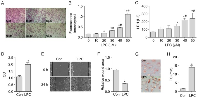
Effects of LPC on the generation of ROS and LDH in HUVE-12 cells, and activation of MPs induced by co-culture with LPC-injured HUVE-12 cells. (A) Representative images of hematoxylin and eosin staining of LPC-injured HUVE-12 cells. The number of stained cells was counted for each group across four areas at 200× magnification and the mean was estimated. (B) LPC (20, 30, 40 and 50 µM) significantly enhanced the fluorescence intensity of each cell in a concentration-dependent manner. (C) LPC (30, 40 and 50 µM) significantly increased the release of LDH in a concentration-dependent manner. LPC (30 µM) effectively increased the (D) cell viability, and decreased the relative wound area of MPs co-cultured with LPC-injured HUVE-12 cells, as shown in (E) images (magnification, ×100) and (F) graph. (G) Representative images of foam cell formation. LPC (30 µM) effectively increased lipid accumulation in MPs (magnification, ×200). (H) LPC (30 µM) effectively increased the TC content. Data are presented as the mean ± standard deviation of three separate experiments. *P<0.05, vs. 0 µM LPC and/or Con (not treated with LPC) group, #P<0.05, vs. 20 µM and/or 30 µM LPC group. LPC, lysophosphatidylcholine; ROS, reactive oxygen species; LDH, lactate dehydrogenase; MPs, macrophages; Con, control; OD, optical density.

