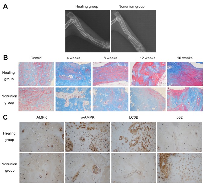
Figure 1.
New bone mineralization and maturation is restrained in the nonunion group, accompanied by decreased AMPK activation and autophagic activity. (A) X-ray image showing the healing status of radial defects 16 weeks following surgery. (B) Masson staining (magnification, ×200) detected osteoid (blue) and mineralized bone (red) in calluses obtained from normal bone (control) and fracture ends 4, 8, 12 and 16 weeks post-surgery. (C) Immunohistochemical staining of AMPK, p-AMPK and autophagy-associated markers (LC3B and p62) 4 weeks after surgery (magnification, ×400). AMPK, adenosine monophos-phate-activated protein kinase; LC3B, microtubule-associated proteins 1A/1B light chain 3B; p-AMPK, phosphorylated-AMPK.

