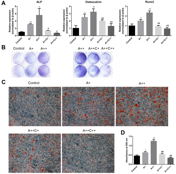
Figure 2.
AMPK activation promotes the differentiation and mineralization of MC3T3-E1 cells. (A and B) Cells were treated with AICAR (A+, 1 μM; ++, 10 μM) and compound C (C+, 0.1 μM; ++, 1 μM) in differentiation medium for 8 days. (A) ALP, osteocalcin and Runx2 mRNA expression were detected by reverse transcription-quantitative polymerase chain reaction. (B) 5-Bromo-4-chloro-3-indolyl phosphate/nitro blue tetrazolium staining was used to determine ALP activity. (C) Cells were cultured for 21 days; representative images (magnification, ×100) of mineralized nodules stained by Alizarin red staining are presented. (D) Quantification of Alizarin red staining with cetylpyridinium chloride. Absorbance was measured at 550 nm. *P<0.05, **P<0.01 compared with the control group; #P<0.05, ##P<0.01 compared with the A++ group. AICAR, 5-aminoimdazole-4-carboxamide-1-β-D-ribofuranoside; ALP, alkaline phosphatase; AMPK, adenosine monophosphate-activated protein kinase; Rnx2, runt-related transcription factor 2.

