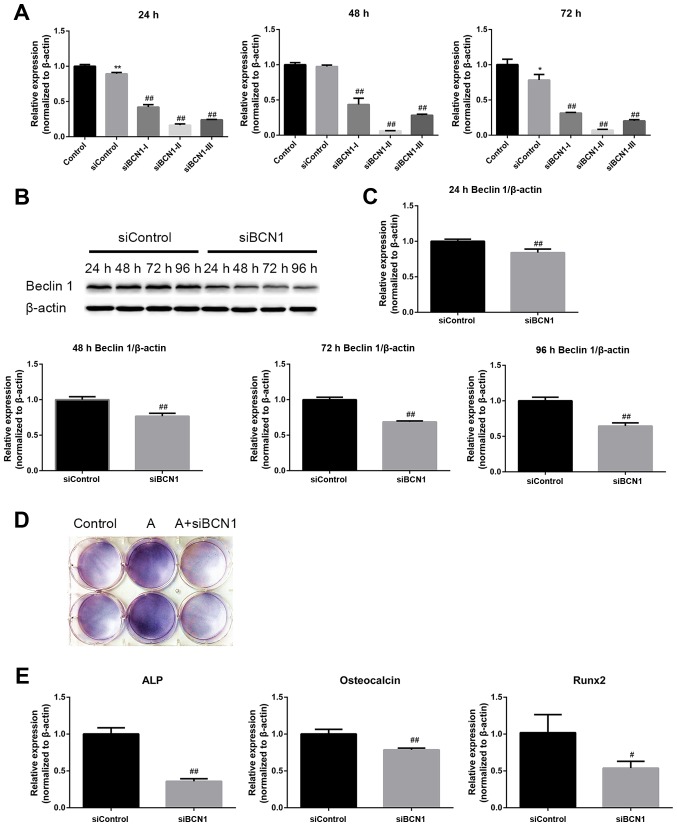
Figure 5.
AMPK activation enhances differentiation of MC3T3-E1 cells via the induction of autophagy. (A) Cells were transfected with the indicated siRNAs, and the mRNA expression levels of BCN1 were measured by RT-qPCR 24, 48 and 72 h post-transfection to assess the silencing effect; siBCN1-II was the most efficient siRNA and was used for further study. (B) Cells were transfected with siBCN1-II and siControl, western blot analyis was performed using an antibody against BCN1 24, 48, 72 and 96 h post-transfection, in order to verify the silencing effect. (C) Semi-quantitative results of western blot analysis. (D and E) A total of 24 h post-transfection with siBCN1-II or siControl, differentiation medium was added and cells were treated with or without AICAR (10 μM) for 8 days. (D) 5-Bromo-4-chloro-3-indolyl phosphate/nitro blue tetrazolium staining was used to detect ALP activity. (E) ALP, OCN and Runx2 mRNA expression was detected by RT-PCR. siControl cells were treated with AICAR. *P<0.05, **P<0.01 compared with the control group; #P<0.05, ##P<0.01 compared with the siControl group. AICAR, 5-aminoimdazole-4-carboxamide-1-β-D-ribofuranoside; ALP, alkaline phosphatase; AMPK, adenosine monophosphate-activated protein kinase; BCN1, Beclin 1; OCN, osteocalcin; Runx2, runt-related transcription factor 2; si/siRNA, small interfering RNA.

