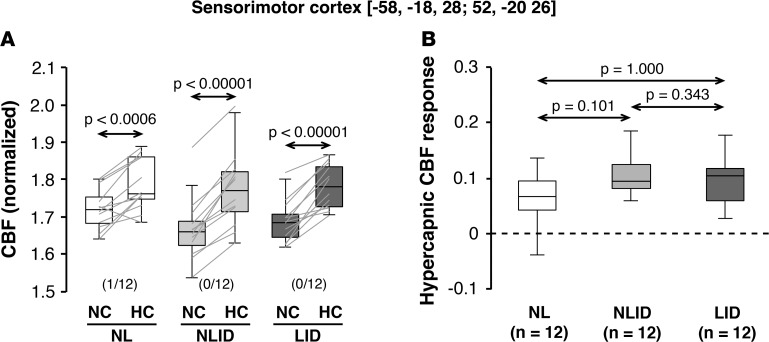
Figure 4. Hypercapnic CBF responses in the sensorimotor cortex.
Globally normalized cerebral blood flow (CBF) was measured in the sensorimotor cortex (SMC) region in which baseline (off-state) metabolic activity was previously found to be elevated in levodopa-induced dyskinesia (LID) subjects (n = 12) compared with their non-LID (NLID) counterparts (n = 12) (see Results). (A) Hypercapnic CBF increases in this region were observed consistently in LID, NLID, and normal (NL) subjects (n = 12). Horizontal arrows denote significance levels (P values) for each group according to the paired Student’s t test. (B) In contrast to the putamen (Figures 1 and 3), hypercapnic responses in this region did not differ across the three groups of subjects. Horizontal arrows denote significance levels (P values) for group comparisons according to the post-hoc Bonferroni test of 1-way ANOVA. (Graphical displays as in Figures 1–3.)

