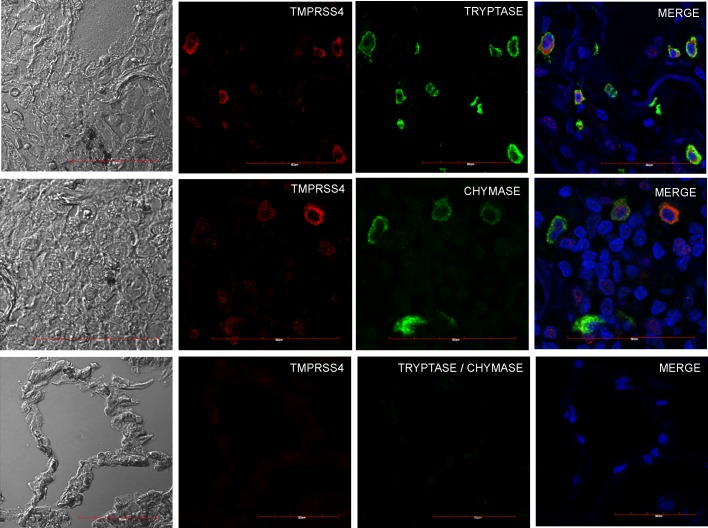
Fig 3. Co-localization of TMPRSS4, tryptase and chymase in IPF and control lungs.
Representative images of immunofluorescence staining performed with specific antibodies against each molecule in lung tissue sections from IPF patients. Top and central row: IPF tissues coexpressing TMPRSS4 and tryptase or chymase; bottom row: normal lung showing no expression. Tissues were stained for TMPRSS4 (Dylight-549, red), tryptase (AF-488, green upper panel) and chymase (AF-647, green middle panel). The colocalization of TMPRSS4 and tryptase or chymase was determined by fluorescence microscopy and images were merged to resolve the co-localization of these proteins. Co-localization of TMPRSS4 was observed with both triptase and chymase. Data are representative of three independent experiments.

