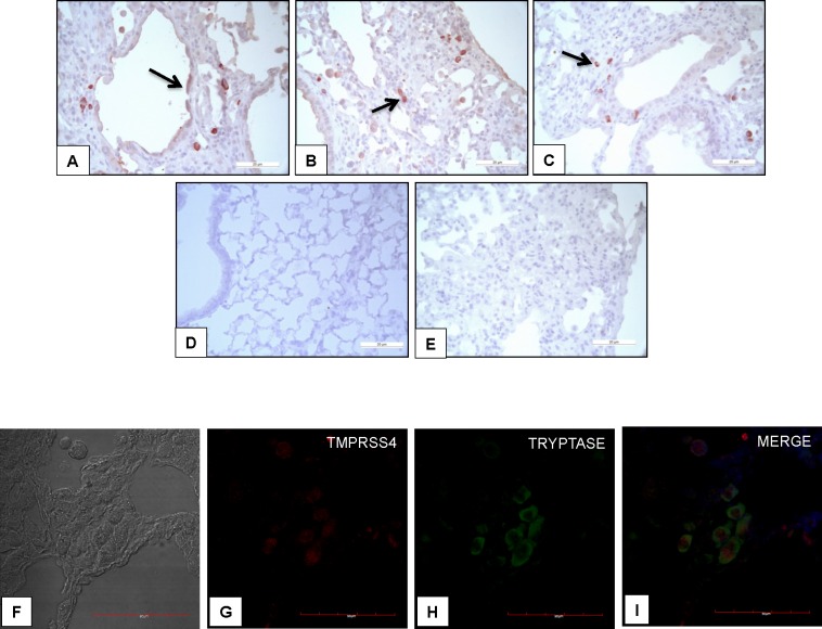
Fig 9. TMPRSS4 is expressed by epithelial and mast cells in bleomycin-induced pulmonary fibrosis.
Representative photomicrographs of immunohistochemical staining performed with antibody against TMPRSS4 in lung tissue sections from WT mice injured with bleomycin. The immunoreactive enzyme was observed in epithelial (panel A, black arrow) and interstitial cells (panels B and C black arrows). Panel D: no positive staining was detected in normal lungs. Panel E: shows the negative control where the specific antibody was omitted. All sections were counterstained with hematoxylin. Panels F-I: Representative images of immunofluorescence staining performed with specific antibodies against TMPRSS4 and tryptase; Tissues were stained for TMPRSS4 (Dylight-549, red) and tryptase (AF-488, green). The colocalization of TMPRSS4 and tryptase was determined by fluorescence microscopy and images were merged to resolve the co-localization of these proteins.

