
Figure 5.
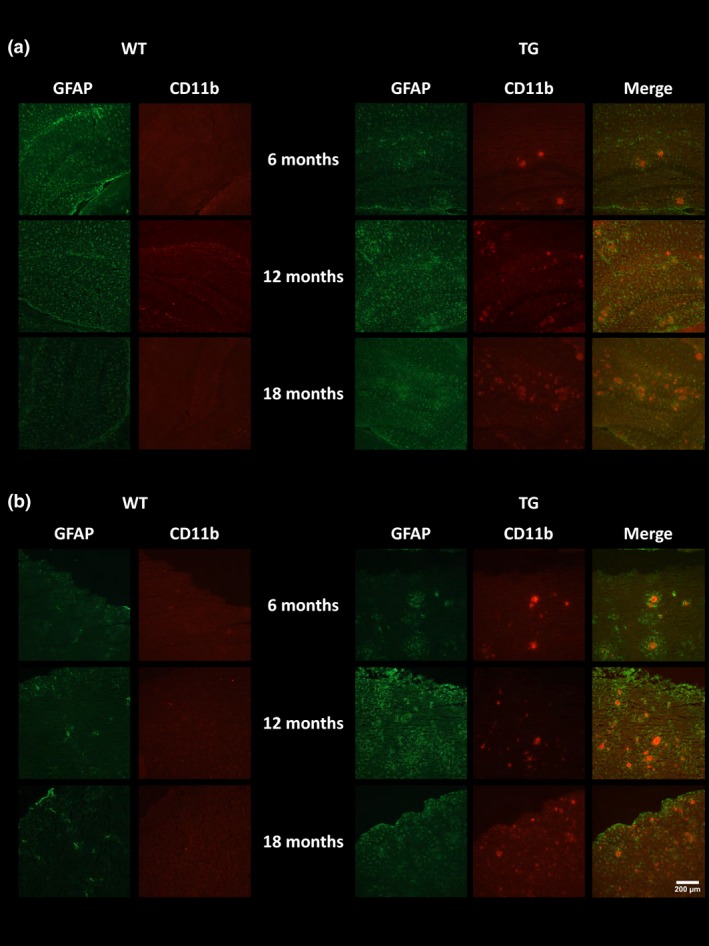
Immunoreactivity of glial fibrillary acidic protein (GFAP) (green) and CD11b (red). Representative images of double staining in the hippocampus (a) and cortex (b) of WT and APP swe×PS1Δe9 mice at 6, 12 and 18 months of age. Pictures were taken at 10 × magnification between bregma −2.06 mm and −2.30 mm. Scale bar represents 200 μm.