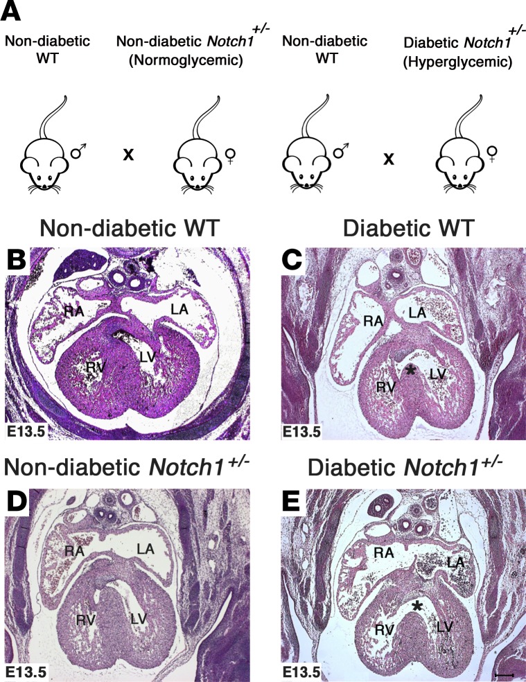
Figure 2. Exposure to maternal diabetes and Notch1 haploinsufficiency leads to increased incidence of ventricular septal defects.
(A) Breeding scheme demonstrating nondiabetic WT males crossed with nondiabetic (normoglycemic) and diabetic (hyperglycemic) Notch1+/– females. (B–E) Representative H&E-stained images showing the location of ventricular septal defects (*) in E13.5 WT and Notch1+/– hearts exposed to maternal diabetes as compared with nondiabetic WT and Notch1+/– controls. RA, right atrium, LA, left atrium; RV, right ventricle; LV, left ventricle. Scale bar: 200 μm.

