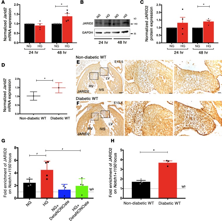
Figure 4. Jarid2 mediated regulation of Notch1 with hyperglycemia.
(A) RT-qPCR and (B) representative immunoblot showing Jarid2 transcript and JARID2 protein levels in NG- (5.5 mM) and HG-treated (25 mM) AVM cells 24 and 48 hours after treatment (n = 4; mean ± SEM; *P < 0.05, 2-tailed Student’s t test). (C) Quantification of relative expression normalized to GAPDH (n = 4; mean ± SEM; *P < 0.05, 2-tailed Student’s t test). (D) Examination of E13.5 murine hearts (n = 6 hearts pooled together/group) exposed to nondiabetic and diabetic environments shows upregulation of Jarid2 mRNA by RT-qPCR (mean ± SD; *P < 0.05, 2-tailed Student’s t test). (E and F) Increased JARID2 protein in embryonic hearts exposed to maternal diabetes by immunohistochemistry (n = 3). Square boxes in E and F are shown in higher magnification from left to right. LV, left ventricle; RV, right ventricle; IVS, interventricular septum. Scale bars: 100 μm (E and F, left column); 20 μm (E and F, center and right columns). (G) ChIP-qPCR on AVM cells in NG or HG revealed enrichment of JARID2 on Notch1+1150 locus with HG and lack of enrichment with addition of 250 μM DetaNONOate (n = 4; mean ± SEM; #P < 0.05, 2-tailed Student’s t test, not significant with Holm-Bonferroni correction). (H) In vivo ChIP-qPCR on E13.5 hearts exposed to nondiabetic and diabetic environments (n = 3). Rabbit IgG served as mock control, shown as dotted line (set to 1) (mean ± SEM; *P < 0.05, 2-tailed Student’s t test).

