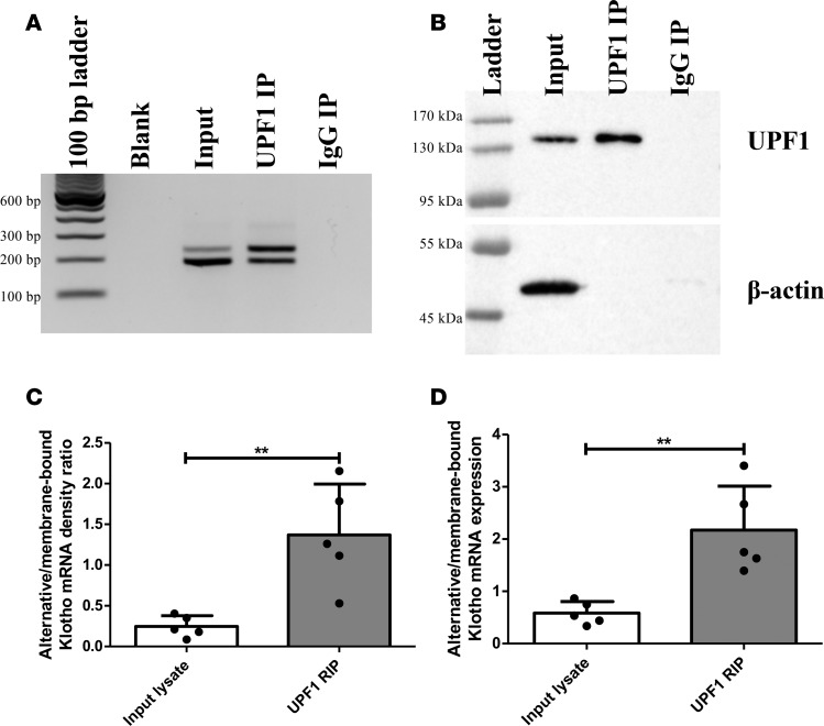
Figure 3. RNA IP of HK-2 cell lysate reveals enrichment of the alternative Klotho mRNA associated with UPF1.
(A) RT-PCR analysis for both Klotho transcripts in HK-2 cell lysate (input), after IP for UPF1, and after IP with rabbit IgG, showing enrichment for the alternative Klotho mRNA in UPF1-associated RNA. (B) Western blot analysis for UPF1 and β-actin in HK-2 cell lysate and after IP with rabbit anti-UPF1 or rabbit IgG, showing enrichment for UPF1 and depletion of other proteins, as indicated by absence of β-actin in IP fractions. (C) Densitometric quantification of A, expressed as the ratio of the alternative and membrane-bound Klotho mRNAs. (D) qPCR analysis for both Klotho transcripts, using ΔCt = Ct(alternative Klotho transcript) – Ct(membrane-bound Klotho transcript), confirms the RT-PCR results. **P < 0.01, as tested by Student’s t test. All individual data points represent values from independent experiments (with mean ± SD).

