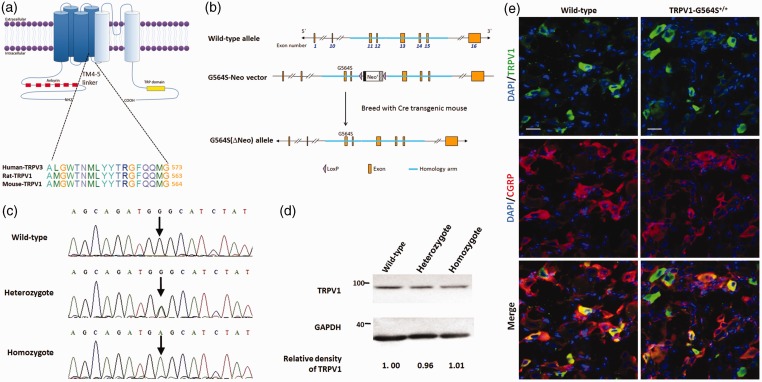
Figure 1.
Generation and characterization of TRPV1 G564S knock-in mice. (a) Topology of the TRPV1 channel subunit and homology between TRPV3 and TRPV1; (b) schematic of the domain structure of (upper) mouse Trpv1 genomic loci, (middle) the construct showing fusion of the homologous arm containing G564S in exon 11 followed by the neo selection marker flanked by loxP sites, and (lower) a map of the G564S (ΔNeo) allele; (c) gene sequence analysis identifying the genotype of a wild-type animal (upper), heterozygote (middle), and homozygote (lower); (d) representative Western blots of DRG tissue showing the comparative expression levels of TRPV1 in wild-type, heterozygotic, and homozygotic mice after normalizing to β-actin. Note the comparable relative densities of TRPV1 blots scaled to the wild type; (e) representative immunofluorescence staining of DRG sections revealing the comparable distribution of TRPV1 and colocalization with CGRP in TRPV1 G564S+/+ mice (right) and wild-type controls (left). Scale bars, 20 μm. TRPV: transient receptor potential vanilloid; GAPDH: glyceraldehyde-3-phosphate dehydrogenase; DAPI: 4', 6-diamidino-2-phenylindole; CGRP: calcitonin gene-related peptide.

