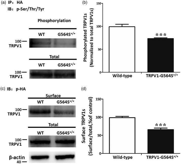
Figure 6.
Reduced phosphorylation and membrane localization of TRPV1 channels caused by G564S mutation. (a) Western blots showing the phosphorylated TRPV1 protein levels in HEK293a cells transfected with HA-TRPV1 or HA-TRPV1-G564S plasmids after immunoprecipitation by anti-HA antibody. (b) Analysis showing a significantly lower phosphorylation level of the TRPV1 G564S channel than the wild type control. Error bars, SEM; ***p < 0.001, unpaired Student’s t test. All experiments were repeated at least four times. (c) Surface biotinylation assays for the amount of TRPV1 at the cell surface. (d) Analysis showing the ratio of surface to total TRPV1 normalized to the wild-type control. Error bars, SEM; ***p < 0.001, unpaired Student’s t test. All experiments were repeated at least four times. TRPV: transient receptor potential vanilloid; HA: hemagglutinin; WT: wild type.

