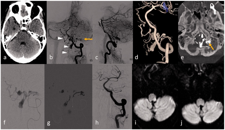
Figure 1.
(a) Non-contrast computed tomography (NCCT) of the head showing focal subarachnoid haemorrhage (SAH) in the left cerebello-pontine angle. (b), (c) Left vertebral artery angiogram antero-posterior and lateral views revealed dural arteriovenous fistula (AVF) (arrow) in the left lateral aspect of the foramen magnum fed by the posterior meningeal branch of the left V3 vertebral artery and venous drainage through the medulla bridging vein with reflux into the contralateral anterior medullary-anterior pontomesencephalic (AM-APM) venous system (arrowhead) and anterior spinal vein (arrowhead). (d) Three-dimensional (3D) image depicts better anatomy of the dural AVF; (e) multiplanar reconstruction (MPR) reformatted from the 3D source image shows the exact site of the fistula at the left posterior condylar canal (arrow) with venous pouch at the dorsal aspect of the foramen magnum. (f) Microcatheter injection through the feeding artery and (g) glue cast in the fistula and also into the draining venous pouches are shown. (h) Control angiogram through the left vertebral artery revealed complete obliteration of the AVF. Diffusion-weighted imaging (DWI) revealed focal restricted diffusion along the left dorso-lateral medulla (i), resolved in the subsequent DWI image (j) performed after 10 days.

