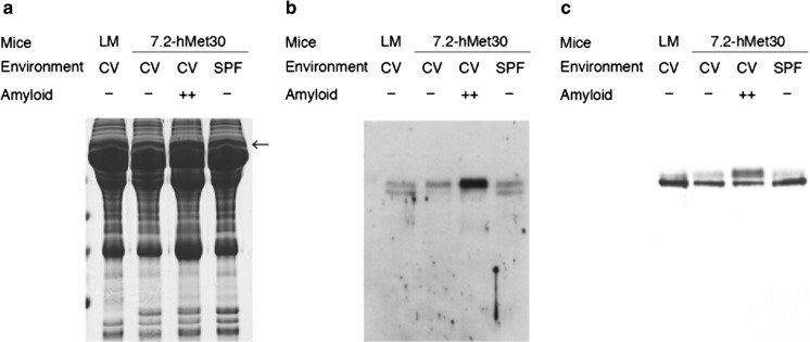
Fig. 1.
Comprehensive analysis of serum proteins in amyloid-positive and amyloid-negative 7.2-hMet30 mice. Differences in amyloid-positive serum were found in the band pointed by an arrow by means of proteomic analysis after SDS-PAGE. HPX and Tf were detected by MS analysis and they were found only in the amyloid positive serum in the band with an arrow (a). The presence of the protein differences detected, HPX (b) and Tf (c), was confirmed by using western blotting. LM, littermate

