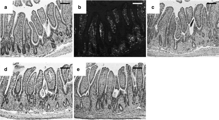
Fig. 2.
Histochemical and immunohistochemical analyses of amyloid deposits in the small intestine of a 7.2-hMet30 mouse kept under CV conditions. Congo red-positive deposits were present in the lamina propria of the small intestine (a). These deposits showed apple green birefringence under polarized light (b). Amyloid deposits demonstrated positive reactions with anti-human TTR antibody in our immunohistochemical analysis (c). Immunohistochemical analysis suggested co-precipitation of HPX in TTR amyloid deposits (d), and the presence of Tf (e). Bars = 100 μm. (Color figure online)

