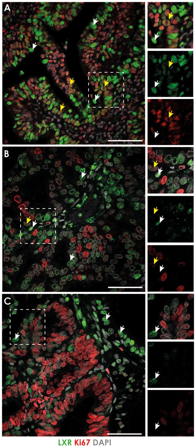
Figure 2.

Expression of LXR and the proliferation marker Ki67 in endometrial cancer. The expression of LXR (antibody identified both isoforms) and the proliferation marker Ki67 was assessed by immunohistochemistry in endometrial cancer tissue sections. In well-differentiated cancers (A), LXR was expressed throughout the tissue and localised to the nuclei of both stromal and epithelial cells (green staining). Nuclear immunoexpression of Ki67 (red staining) was detected and co-localised with LXR expression (yellow arrows) although some LXR-positive cells did not co-express Ki67 (white arrows). In moderately differentiated cancers (B) both markers were detected but did not appear to co-localise; only few cells expressed both LXR and Ki67 (yellow arrows). Most LXR-positive cells did not co-express Ki67 (white arrows). This was also true of poorly differentiated cancers (C), few cells expressed both LXR and Ki67 (yellow arrows) although LXR-positive cells were found in close association with proliferating cells (white arrows). Images representative of at least 3 different patients per cancer grade. Nuclear counterstain DAPI (grey). All scale bars 50 µM.

 This work is licensed under a