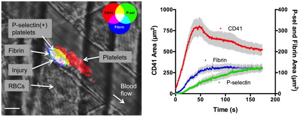
Figure 2. Spatio-temporal regulation of platelet activation following vascular injury.
A) Photomicrograph shows platelet accumulation, activation and fibrin deposition following laser-induced injury in a mouse cremaster arteriole. Note the leakage of RBCs into the extravascular space as a result of the injury, and the spatial localization of P-selectin positive platelets adjacent to the site of injury. Scale bar is 10 μm. B) Graph depicts typical kinetics of platelet accumulation, activation and fibrin deposition following laser-induced injury in cremaster arterioles of C57Bl/6 mice. Platelet accumulation is shown in red (left axis), P-selectin in green and fibrin in blue (right axis). Values are mean±SEM for n=27 thrombi.

