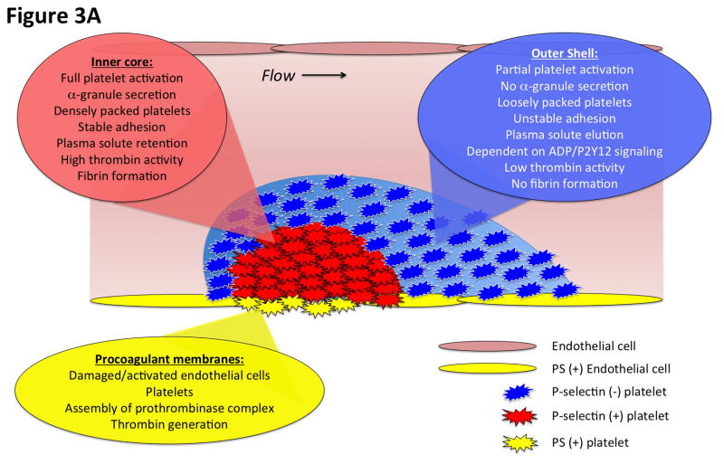
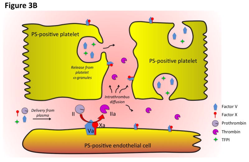
Figure 3. Diagrammatic representation of the spatial distribution of coagulation events and platelet activation at the cellular and molecular levels.
A) This diagram illustrates the heterogeneity of platelet activation at a site of vascular injury. Two regions, an inner core and an outer shell, may be defined based on a number of characteristics including platelet activation state, platelet packing density, porosity and level of thrombin activity. Localization of procoagulant surfaces and fibrin formation is also shown. B) This diagram illustrates coagulation events at the molecular level and how they are regulated by the local microenvironment. Assembly of the prothrombinase complex on PS-positive endothelial cell and platelet membranes localized at the site of injury leads to thrombin generation. The spatio-temporal distribution of thrombin activity is determined by its sites of generation, localization of inhibitors, and physical factors in the local microenvironment that regulate molecular transport. Molecular transport phenomena also regulate the delivery of additional pro- and anti-coagulant factors from the bulk flow and those released locally from platelet granules (e.g. factor V and TFPI).


