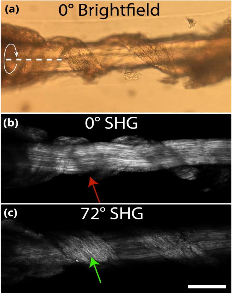
Fig. 5.
(a) Brightfield and (b) SHG images of coiled tendon oriented at 0°; (c) SHG image of tendon rotated 72°. The red arrow indicates missing fibers due to orientation parallel to excitation and the green arrow shows fibers reappearing after the 72° sample rotation. The curved arrow and dashed line delineate the axis of rotation. Scale bar = 100 µm.

