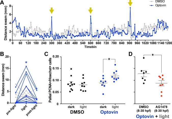
Figure 7. Photoactivation of DRGs using Optovin increases pallial neurogenesis.
5 dpf larvae incubated in Optovin exhibit repeatable movement bursts in response to light (A). Points on each line represent the mean distance swam (n = 12) during 5 s time bins by larvae treated with either Optovin (blue) or DMSO (grey) and exposed to 2 s of light (yellow arrows) every 5 min. (B) Expanded view of first light presentation from (A), including the 5 s of darkness prior to light presentation (pre-light), the 5 s time bin including light presentation for the first 2 s (light), and the following 5 s of darkness (post-light). Treating 5 dpf larvae immobilized in agarose with 2 s light pulses every 5 min for 5 hr increased the number of PCNA+ cells in the pallium 12 hr post treatment only when larvae were incubated with Optovin (C; DMSO dark n = 10, DMSO light n = 9, AG1478 dark n = 8, AG1478 light n = 9). Larvae treated with AG1478 from 8 to 30 hpf failed to exhibit an increase in the proportion of PCNA +cells in the pallium following this same Optovin + light treatment compared to controls (DMSO control n = 7, AG1478 n = 8). *p<0.05. Data are represented as mean ± SEM.
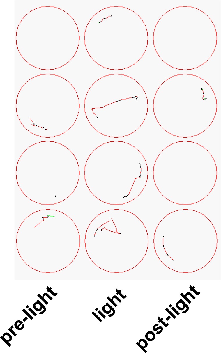
Figure 7—figure supplement 1. Example movement traces from 5 s timebins in 4 wells of larvae incubated with 10 µM Optovin prior to, during, and following a 2 s presentation of white light (800 lux).


