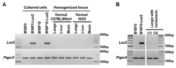
Fig. 1.
Specific amplification of Luc2 and Ptger2 gene fragments with genomic DNA from mouse cells and organs. 100 ng of genomic DNA was used as template in each 25μl reaction and PCR was performed for 30 cycles with primer pairs as described in Experimental section. The products were resolved on 2.5% agarose gel. (A) Amplification of Ptger2 but not Luc2 in genomic DNA from normal mouse organs (lungs, liver and brain). (B) Amplification of both Ptger2 and Luc2 in genomic DNA from mouse lungs with Luc2 positive metastatic melanoma. Sample C1 and C5 represent individual animals described in Table 2, in which Luc2 positive cells were detected in these two mice using bioluminescence imaging.

