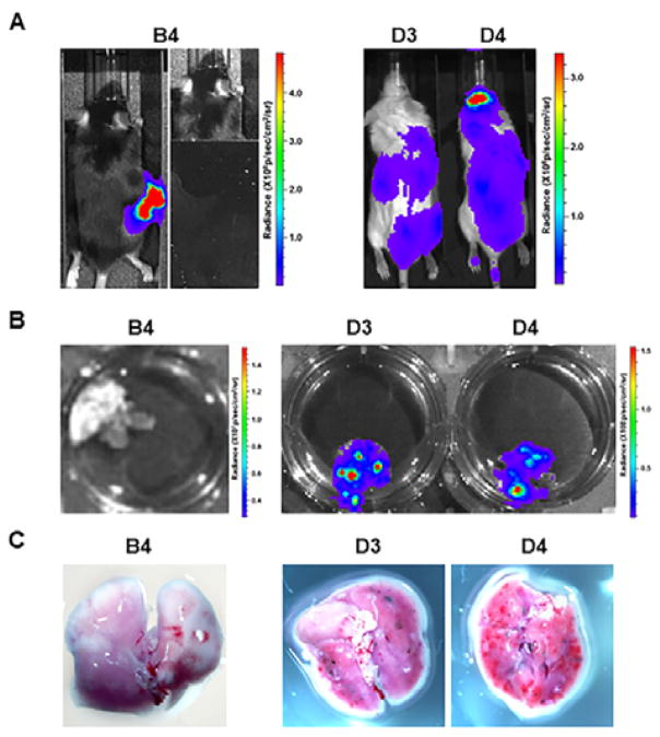
Fig. 4.
Comparison of spontaneous and experimental lung metastasis detection with bioluminescence imaging and close-up photography. Summaries of representative mice for spontaneous metastasis (B4) and experimental metastasis (D3 and D4) were listed in Table 2. (A) In vivo bioluminescence imaging one day before the mice were terminated as listed in Table 2. On the right side of panel B4, the primary melanoma was shielded for better detection of lung metastases. (B) Ex vivo bioluminescence lung imaging. (C) Photos for black B16F10-Luc2 macrometastases on lung surfaces.

