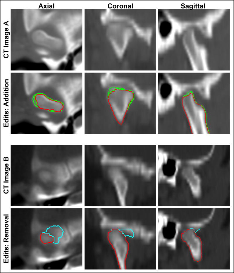
Figure 3.
Examples of post-processing addition and removal of voxels for under-segmented and over-segmented regions displayed in all three orientations. Top panel: CT image A displays the temporomandibular joint region of a male case (13 years 10 months old). Red represents the automatically segmented condyle region while green represents the voxel additions edited manually by a trained researcher. Lower panel: CT image B displays the temporomandibular joint region of another male case (2 years and 5 months old). Cyan represents the over-segmented region, originally segmented automatically, but edited for removal manually by a trained researcher.

