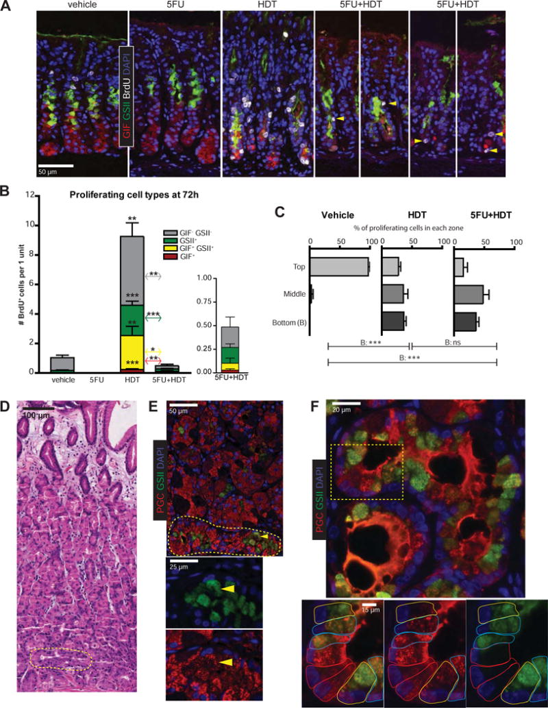
Figure 2. Metaplastic cells arise below the isthmus.

A) Immunofluorescence of stomachs after 72h vehicle, 5FU, HDT, or 5FU+HDT (green, GSII; red, GIF; white, BrdU; blue, DAPI; arrowheads, rare BrdU+ cells in 5FU+HDT). B) Proliferating cell populations quantified. C) Location of BrdU+ cells below the bottom-most AAA+ pit cell. D) Hematoxylin & eosin of early human SPEM (yellow circle) E) Immunofluorescence on serial section from 2D (red, PGC; green, GSII; blue, DAPI; arrowhead: SPEM cell). Yellow circle is SPEM area. F) Human SPEM within gland base showing transitional ZC➔SPEM forms. Dotted box indicates area shown at higher magnification. Cells outlined in colors according to cell type (red, normal ZCs; blue, hybrid SPEM; yellow, full SPEM). *p≤0.05 **p≤0.01 ***p≤0.001, variance analyzed with ANOVA/Tukey (N ≥ 3 mice/group)
