Figure 2.
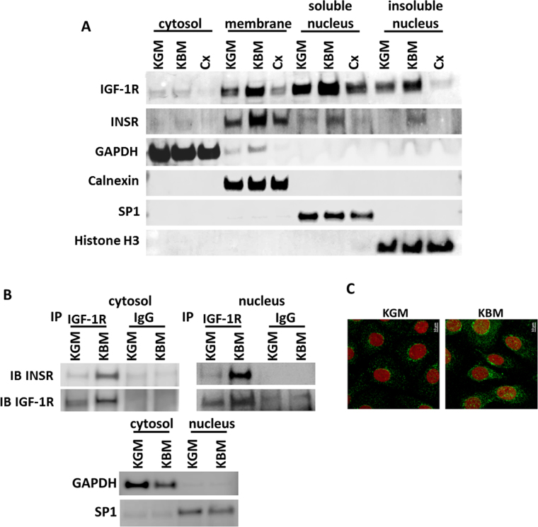
Nuclear accumulation of the IGF-1/insulin Hybrid R occurs de novo in response to growth factor deprivation. (A) hTCEpi cells were cultured in growth, basal or basal media supplemented with 1 μg/ml cycloheximide (Cx) overnight. Cells were lysed and fractionated into cytosolic, membrane, soluble and insoluble nuclear fractions. Consistent with whole cell lysates, immunoblotting for IGF-1R showed an increase in IGF-1R expression in basal media compared to controls. Treatment with cycloheximide blunted the basal increase and reduced expression levels to below those seen in growth media. There was a large increase in nuclear accumulation of IGF-1R in both the soluble and insoluble fractions. Similar to IGF-1R, INSR was also increased in basal media. Cycloheximide treatment of samples in basal media blocked nuclear accumulation of INSR. Immunoblotting for GAPDH, Calnexin, SP1 and Histone H3 were used for cytosolic, membrane, soluble nucleus, and insoluble nucleus fractionation controls. (B) hTCEpi cells were cultured overnight in basal or growth media and fractionated into nuclear and non-nuclear (cytosolic) components. Samples were immunoprecipated for IGF-1R and immunoblotted for INSR and IGF-1R to confirm the presence of nuclear Hybrid-R. There was an increase in Hybrid-R expression in the nuclear compartment in basal media. GAPDH and SP1 were used for cytosolic and nuclear fraction contamination controls. (C) Immunofluorescence for IGF-1R (green) and nuclei (red) shows an increase in IGF-1R expression and nuclear accumulation in basal media compared to growth media.

