Figure 3.
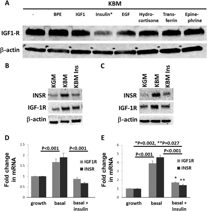
Insulin treatment in basal media restores IGF-1R and INSR to normal levels. (A) hTCEpi cells were cultured in basal media and supplemented individually with each component of the bullet kit (BPE, hEGF, insulin, hydrocortisone, transferrin, epinephrine) and also with 100 ng/ml of IGF-1. Expression of IGF-1R was assessed by immunoblotting. Insulin, a component of the bullet kit, was the only growth factor that decreased expression of IGF-1R. (B) hTCEpi cells were cultured in growth, basal or basal media supplemented with 10 μg/ml insulin. For both receptors, immunoblotting showed an increase in basal media that was attenuated following treatment with insulin. (C) HCECs were also cultured overnight in growth, basal or basal media supplemented with 10 μg/ml insulin. Similar to hTCEpi cells, there was a large increase in INSR and IGF-1R expression in basal media that was reduced following the addition of insulin to the culture media. (D) Real time PCR for IGF-1R and INSR in hTCEpi cells showed that changes in receptor expression levels paralleled mRNA levels with a significant increase in IGF-1R and INSR mRNA in basal media compared to growth media or basal media supplemented with insulin (P < 0.001, Two-way ANOVA, Tukey multiple comparison test). (E) Real time PCR for IGF-1R and INSR in HCECs. There was a significant increase in IGF-1R and INSR in basal media compared to growth conditions or basal media supplemented with insulin (P < 0.001, Two-way ANOVA, Tukey multiple comparison test). There was a small, but significant difference between IGF-1R and INSR mRNA in basal media supplemented with insulin compared to the growth condition (*P = 0.002 for IGF-1R and **P = 0.027 for INSR, Two-way ANOVA, Tukey multiple comparison test). Data represented as mean ± standard deviation of fold change compared to growth condition.

