Figure 7.
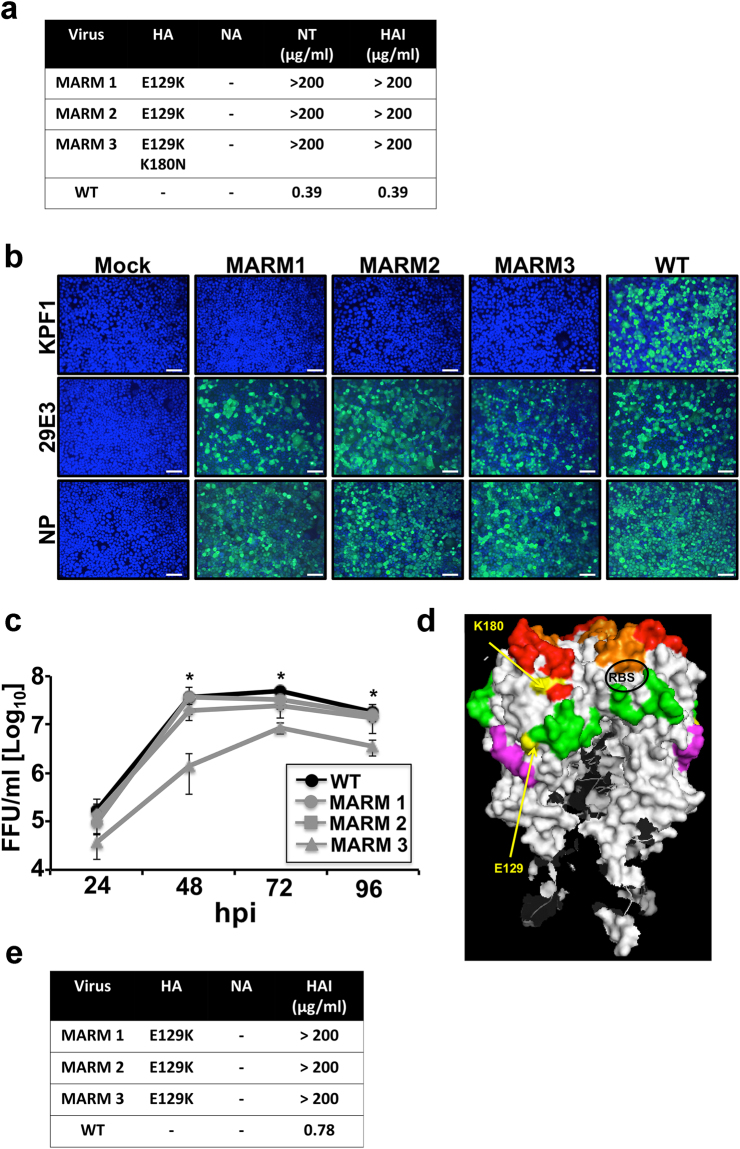
Generation and characterization of MARMs. (a) Amino acid mutations in the HA and NA of pH1N1 WT or mAb-resistant mutants (MARMs 1, 2 and 3) after 5 rounds of selection in the presence (MARMs) or absence (WT) of hmAb KPF1. The mutations effects on reactivity with the hmAb KPF1 were also evaluated in a microneutralization assay (NT50) and HAI. (b) Characterization of MARMs of pH1N1 by immunofluorescence. MDCK cells were mock infected (Mock) or infected (MOI 0.01) with pH1N1 WT or the MARMs (1, 2 and 3). At 36 h p.i., cells were fixed and protein expression was evaluated by IFA using the hmAb KPF1, or the mouse mAbs 29E3 (anti-HA) and HB-65 (anti-NP). DAPI was used for nuclear staining. Merge from representative images (10x magnification) are included. Scale bar, 50 nm. (c) Multicycle growth kinetics of pH1N1 WT and MARMs in MDCK cells. Virus titers in TCS of MDCK cells infected (MOI, 0.001) with pH1N1 WT or MARMs viruses were analyzed at the indicated h p.i by immunofocus assay (FFU/ml) using the anti-NP mouse mAb HB-65. Data represent the means ± SDs of the results determined for triplicate wells. *Indicates p < 0.05 (WT versus MARM 3) using a one-tailed Student’s t test. (d) Tridimensional protein structure for the globular head of HA of pH1N1. The image was created using the software program PyMol and the published structure for the HA of pH1N1 (3LZG54,). Positions of amino acid substitutions in the MARMs (E129 and K180) are colored in yellow. The residues at each antigenic site are colored as red for the Sa site, orange for the Sb site, green for the Ca site, and magenta for the Cb site. The receptor binding site (RBS) location in the structure is indicated. (e) Generation of MARMs for TX H1N1. Amino acid changes in the HA and NA of TX H1N1 WT or MARMs (1, 2 and 3) after 5 rounds of selection in the presence (MARMs) or absence (WT) of hmAb KPF1. The effect of E129K mutation on reactivity with KPF1 was also evaluated in a HAI assay, using WT TX H1N1 as an internal control.

