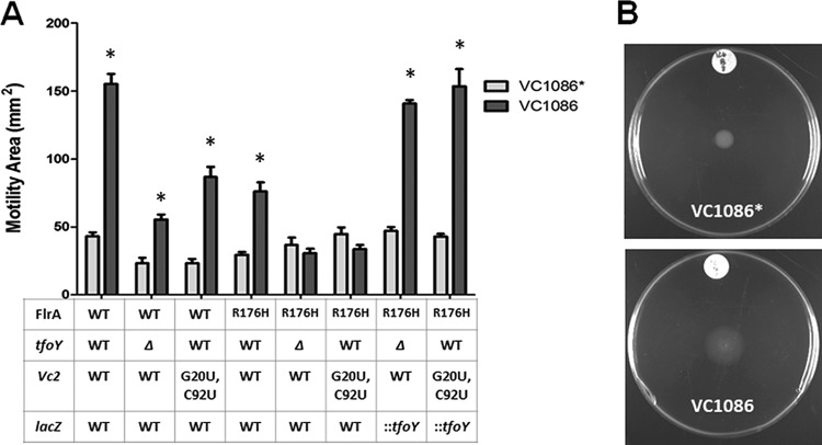
FIG 7.
Inhibition of FlrA and induction of TfoY both promote enhanced motility at the low-c-di-GMP state. (A) Motility of the mutants indicated on the x axis in low-nutrient LB 0.35% agar plates is indicated at 6 h. Dark bars represent the low-c-di-GMP state, while light bars represent the intermediate-c-di-GMP state. All indicated mutations are located on the chromosome. Error bars indicate the standard deviation. *, P < 0.05 in a comparison of VC1086 versus VC1086*. (B) Representative 11-h motility plate images showing dispersive motility at low c-di-GMP concentrations.

