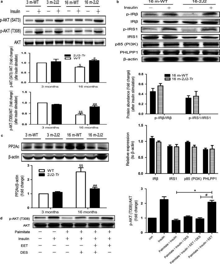
Figure 3.

CYP2J2 overexpression‐mediated improvement in insulin sensitivity coincides with repressed PP2A in liver. Mice were anesthetized after overnight fasting, and 1 IU per kg insulin or an equal volume of vehicle was administered through the portal vein. Liver tissue was collected 120 s after the injection and analyzed by Western blot. (a) Immunoblots of p‐Akt (S473), p‐Akt (T308), and T‐Akt in liver of 3‐ or 16‐month‐old WT and 2J2‐Tr mice. (b) Liver lysates from 16‐month‐old WT and 2J2‐Tr mice were immunoblotted using the antibodies as shown. (c) Representative immunoblots and quantitation of PP2Ac and β‐actin in liver extracts. Data are shown as means ± SE (n = 6 per group). § p < .05, 16mWT vs. 3 mWT; §§ p < .01, 16 mWT vs. 3 mWT; # p < .05, 16 m2J2‐Tr vs. 16 mWT; ## p < .01, 16 m2J2‐Tr vs. 16 mWT. In addition, HepG2 hepatocytes (ATCC) were exposed to 0.25 mm palmitate for 24 h in the presence or absence of 10 nm D‐erythro‐sphingosine (DES) and 1 μm 14,15‐EET in serum‐free medium prior to stimulation with insulin (Sigma–Aldrich) or vehicle control (20 nm for 10 min). (d) Resulting cell lysates were immunoblotted using p‐Akt (T308) and T‐Akt antibodies. Values presented are the mean ± SE from three independent experiments. *p < .05, # p < .05
