Figure 4.

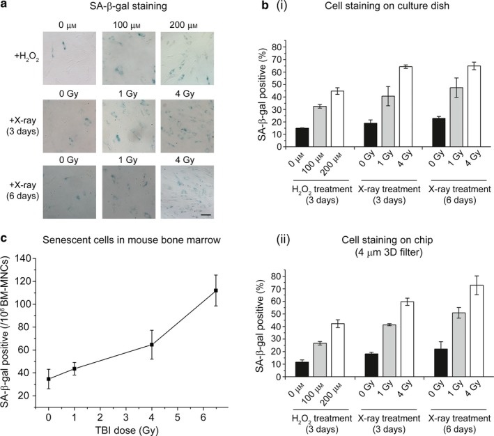
Application of senescence chip for analysis of senescent cells in biofluids. (a) SA‐β‐gal staining of mesenchymal stem cells (MSCs) cultured on a 12‐well plate. The MSCs were treated with different doses of hydrogen peroxide (H2O2, 0, 100, 200 μm) and X‐ray (0, 1, 4 Gy), and analyzed 3 days and 6 days after the treatments. Cells stained blue are SA‐β‐gal positive. SA‐β‐gal: senescence‐associated beta‐galactosidase. (b) Quantitation of SA‐β‐gal staining of MSCs on culture dish (i) and MSCs isolated from human whole blood on the senescence chip (ii). The percentage of SA‐β‐gal positive was calculated for the blue‐stained MSCs among the total MSCs. (c) Isolation and analysis of senescent cells from mouse bone marrow after TBI of 0, 1 Gy, 4 Gy, and 6.5 Gy X‐ray radiation (n = 4), respectively. TBI: Total body irradiation. Scale bar represents 100 μm in (a)
