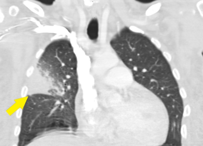
Figure 1.

CT chest with intravenous contrast. There is dense consolidation and enhancement of the right upper lobe, which causes mass effect and bowing of the minor fissure (arrow). There are central areas of low attenuation suggesting necrosis or intrapulmonary abscess, and there is surrounding ground-glass opacity involving the majority of the inferior lateral right upper lobe.
