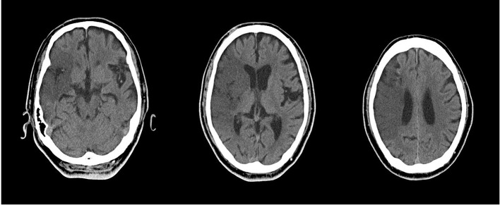
Figure 1.
CT scans (1 day after onset). Three slices (3 cm interval). Extensive cerebral infarction in the middle cerebral artery area due to occlusion from the right internal carotid artery. Old cerebral infarct exists in right and left basal ganglia. In addition, there is contraction in both hippocampi.

