Figure 1.

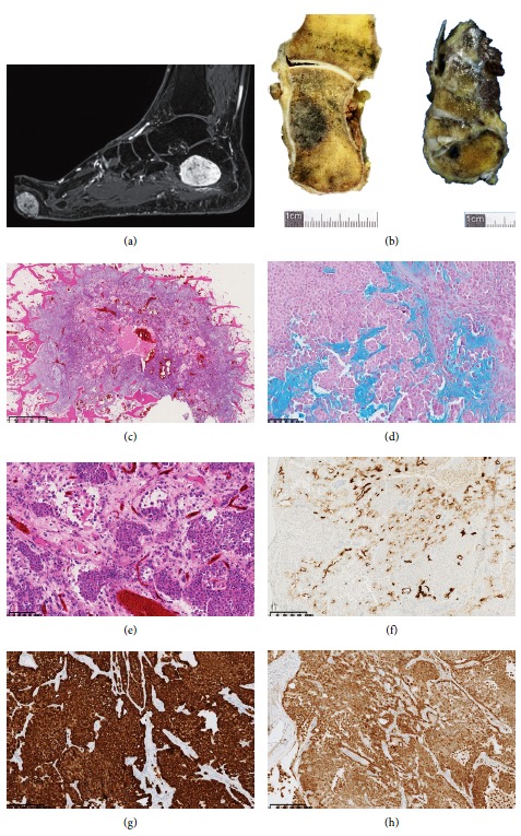
Characterization of the lesions on MRI, macroscopic, microscopic, and immunohistochemical level. (a) MRI gadolinium-enhanced fat-saturated T1-weighted image in the sagittal plane showing an intraosseous lesion of the calcaneus and a plantar soft tissue lesion of the forefoot (arrows). (b) Macroscopic appearance of lesions in the calcaneus and in the cuneiform bone (arrows). (c–e) Lobulated infiltrative pattern made of plasmacytoid cells (H and E). (d) Staining of the chondromyxoid stroma by alcian blue. (f–h) Focal expression of the tumor cells for EMA (f), diffuse expression for cytokeratin AE1/AE3 (g), and diffuse expression for S100 protein (h). Sections of bone were performed using the diamond band saw (EXAKT312, Germany) and decalcified with a formamid solution (DC1, V.W.R.) after formalin fixation. 5 µm thick sections of the paraffin-embedded material were stained with H and E (Symphony 5-Plus, Roche). Immunohistochemistry experiments were performed according to standard procedures. Primary antibodies used on XT benchmark platform (Ventana) were CKAE1/AE3 (cloneAE1/AE3; 1.8 mg/L), EMA (clone E29; 2.4 mg/L), protein S100 (rabbit polyclonal; 1/100), GFAP (rabbit polyclonal; 1/500), alpha-smooth muscle actin (clone 1A4; 0.4 mg/L), desmin (clone D33; 2.05 mg/L), INI1/BAF47 (clone 25/BAF47; 2.5 mg/L), and vimentin (clone V9; 0.5 mg/L).
