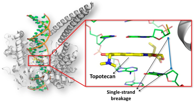
Figure 2.
The poisoned topoisomerase I-DNA complex is shown. The picture has been obtained using the software PyMol version 1.7 (Schrödinger, New York, NY, USA; http://www.pymol.org). The structure of the human topoisomerase is represented in white cartoon, while the DNA double helix is colored in orange (PDB code: 1K4T [121]). In the close-up, the intercalating event of the topoisomerase I poison topotecan (shown in yellow sticks) is reported.

