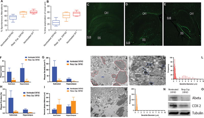
Fig. 2.

Improved hippocampal learning and memory in 5XFAD mice correlates with reduced Aβ expression. A) Assessment of the working memory of three cohorts of animals, including untreated 5XFAD, respiratory curcumin-exposed 5XFAD (12 × 5 mg/Kg) and untreated wt mice (n = 10, each) using a Y-shaped maze (p < 0.0001, one-way ANOVA). B) A two-trial Y-shaped maze was used to test the spatial memory of the animal cohorts noted in (A). The effect of the curcumin after 4 months of inhalation therapy (7 mg/mL, × 12) on spatial memory following 6 min of Y-maze exploration. Each bar represents mean ± SD, statistical significant differences (p < 0.0001, one-way ANOVA). C–E) Thioflavin T staining of sagittal brain sections, particularly in the hippocampus region, dentate gyrus (DG) and subiculum (SUB) of 5-month-old untreated wt mice (C), untreated 5XFAD (D), and 5XFAD mice exposed via the respiratory system (E). F) The number of plaques in the subiculum and hippocampus in untreated 5XFAD versus their respiratory exposed counterparts (p < 0.05). G) Density of plaques defined as number of plaques per mm2 in the subiculum or hippocampus. H) Plaque fraction defined as the plaque occupancy of plaque in a defined area multiplied by 100. I) Distance between plaques in the region of interest. J) Ultrastructural analysis of the CA3 sub region of the hippocampus of the untreated 5XFAD mice. An electron microscopy micrograph of a large Aβ plaque (asterisk). The data also showed that the plaque is surrounded by swollen dystrophic neurites (dotted red lines). K) The dystrophic neurites of the CA3 of untreated mice are filled with autophagic vesicles that contain amorphous and electron-dense materials (dotted blue lines). L) Gaussian distribution curve of two populations of dendrite diameters observed in untreated 5XFAD mice. M) No detectable Aβ plaques were noted in the CA3 region of the curcumin-respiratory-treated 5XFAD mice. N) No swollen neurites were detected, therefore, only one population of neurites exists in a normal physiological size. O) Western blot analysis of A protein and COX2 expression. All tissues were normalized to β -tubulin. (p < 0.05 for A and p < 0.05 for COX2).
