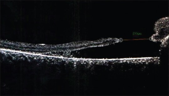
Figure 2.

Optical coherence tomography showing full thickness macular hole at the edge of subretinal hemorrhage (arrowhead). The area of scarring is seen at the nasal edge of the hole (arrow). The minimum linear diameter on optical coherence tomography was 899 μm
