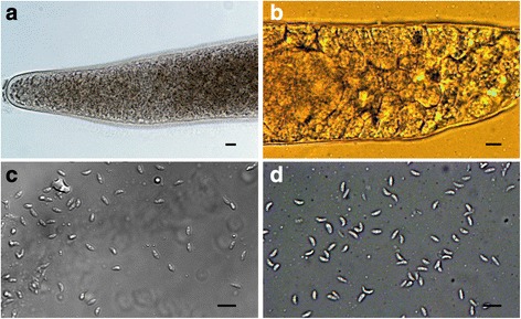
Fig. 1.

Light microscopy photomicrographs of Sarcocystis arctica (a, c) and S. lutrae (b, d) isolated from fresh muscle samples of the red fox (Vulpes vulpes). a Apical portion of sarcocyst; note knob-like villar protrusions on the cyst wall. b Portion of sarcocyst; note septae and thin, smooth wall with the apparent absence of villar protrusions. c, d Banana-shaped bradyzoites released from a broken sarcocysts. Scale-bars: 10 μm
