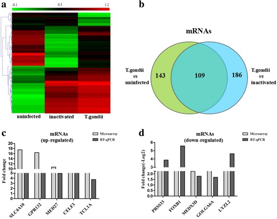
Fig. 2.

Differential expression of mRNAs after T. gondii infection. a The hierarchical clustering of significantly differentially expressed mRNAs (fold change ≥5, P < 0.05) between the uninfected HFF cells (uninfected), the inactivated T. gondii-infected HFF cells (inactivated) and the T. gondii-infected HFF cells (T. gondii). In the heat map, red indicates high relative expression, and green indicates low relative expression. b Venn diagrams indicate the number of total and overlapping mRNAs with significant differential expression in the T. gondii group compared with the uninfected and inactivated groups, respectively. c and d Real-time quantitative PCR (RT-qPCR) validation of the upregulated and downregulated mRNAs (fold change ≥5, P < 0.05) from the microarray data
