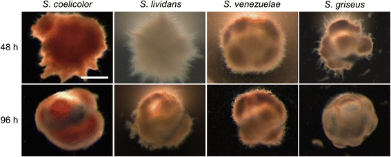
Fig. 1.

Morphology of encapsulated streptomycetes in NMMPmod medium. Microscopy images of microcapsules of Streptomyces coelicolor, Streptomyces lividans, Streptomyces venezuelae and Streptomyces griseus grown in NMMPmod medium at 48 (top panel) and 96 h (lower panel). The scale bar corresponds to 200 μm
