Fig. 1.
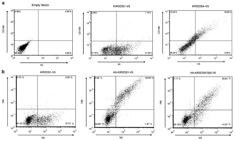
KIR2DS4 and KIR2DS5 are expressed on the surface of NKL cells. Representative flow cytometric analysis of NKL transfectants expressing C-terminally V5-tagged KIR constructs. Total KIR expression was measured with an FITC-conjugated antibody to the V5 tag after the externally stained cells were fixed and permeabilized (X axis). a Representative scatter plots of vector without a KIR insert (left), KIR2DS1 (center) and KIR2DS4 (right) surface expression showing that cells expressing KIR2DS4, but not KIR2DS1, were stained by a PE-conjugated KIR2DS4 (CD158i)-specific mAb, clone FES172 (Y axis). Approximately 26% of KIR2DS4-transfected cells were found in the upper right quadrant of the plot on the right compared to approximately 1% in the center plot. b Representative scatter plots of negative control (left), KIR2DS1 (middle) and KIR2DS5 (*002; right) surface expression. PE positive extracellular expression was detected with an antibody to the HA-tagged KIR (Y axis). Approximately 30% (KIR2DS5 (*002) and 40% (KIR2DS1) of the V5 positive cells exhibited HA surface staining. The negative control expressed a V5-tagged KIR2DS1 molecule without an HA tag

