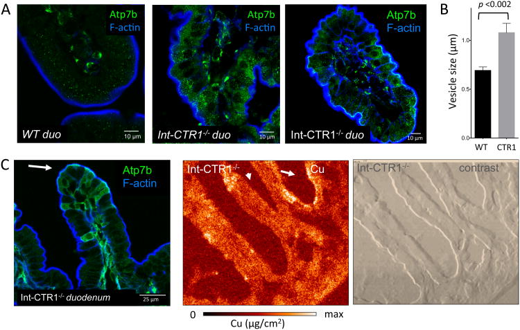
Figure 3. Cu overload correlates with changes in ATP7B distribution in the crypt villus.
(A) ATP7B is highly expressed in the villi of lnt-CTR1-/- enterocytes, which are overloaded with Cu (middle-right), compared with litter mate sex matched controls (left). Under conditions of chronic Cu overload (in the lnt-CTR1-/- tissue) the ATP7B vesicles are significantly more abundant, compared to WT. (B) Quantitation of ATP7B-vesicle size in WT and Int-CTRl-/- mouse duodenum. The lnt-CTR1-/- mouse ATP7B-vesicles are ∼ 60 % larger than those in controls (p-value < 0.002). Ten vesicles were measured from > 10 cells to calculate average vesicle size. (C) ATP7B vesicles are frequently observed (left) on the apical side of the cell (indicated by the white arrow). XFM image of the lnt-CTR1-/- tissue (middle) and phase contrast images illustrating the general morphology of intestine (right). The Cu distribution pattern is altered compared to WT, but still matches that of ATP7B. Cu and ATP7B are accumulated in the villi and are often observed on the apical side of the cell.

