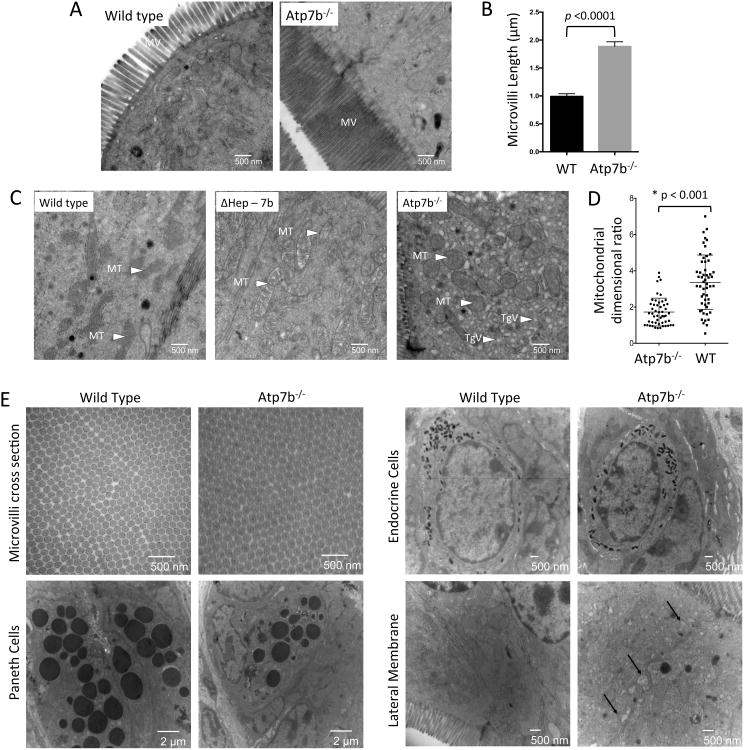
Figure 5. Atp7b-/- enterocytes have altered morphology.
(A) EM micrographs of the brush boarder. The microvilli (MV) of Atp7b-/- absorptive cells are 2 fold longer than controls (n=2 for each genotype). (B) Quantitation of microvilli length in absorptive cells in WT vs Atp7b-/- tissue. (C) In Atp7b-/- cells the mitochondria (MT) are rounded and over populated compared to WT. The Atp7b-/- enterocytes also show accumulation of triglyceride vesicles (TgV) in the cytosol. Intestine from ΔHep-7b mice (with ATP7B specifically deleted in hepatocytes) strongly resembles the WT. (D) Mitochondrial length and width were quantified for >50 mitochondria from >10 cells. The ratio of mitochondrial length-to-width is displayed; values close to 1 represent a spherical morphology. (E) The elongated Atp7b-/- microvilli have no morphological differences in cross-section compared to controls. Paneth cells and endocrine cells do not display morphological changes between wild type and Atp7b-/-. The lateral membrane of Atp7b-/- is altered compared to controls with potential triglyceride accumulation between the membranes (indicated by black arrows).

