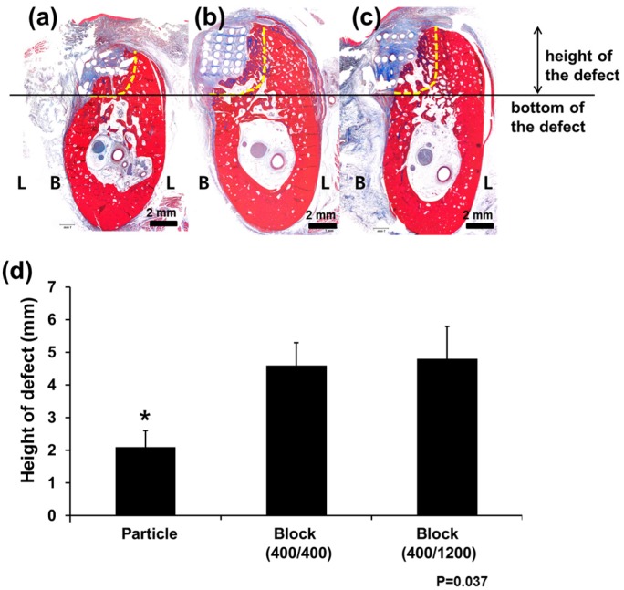
Figure 4.
Histological examination of bone regeneration after PCL scaffold implantation. The (a) Particle group; (b) Block 400/400 group; and (c) Block 400/1200 group; original magnification ×1.25. New bone formation was observed above the bottom of the defects (horizontal line) in all groups. The vertical dimensions of the defects were well-preserved in the (b) Block 400/400 and (c) Block 400/1200 groups, and (d) the difference was statistically significant. “*” indicates statistically significant differences between groups (p = 0.017). L, lingual side; B, buccal side.

