Figure 6.
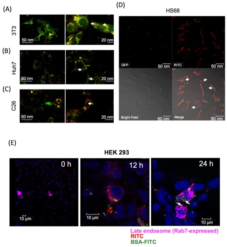
Intracellular protein release from GR-PEI NPs. Confocal laser microscopic images of intracellular protein release. The tomography of cells by confocal laser microscope demonstrated the intracellular BSA-FITC was released from GR-PEI NPs in 3T3 fibroblast cells (A), Huh7 hepatoma cells (B), C26 colon adenocarcinoma cells (C). GFP was released from HS68 fibroblast cells (D). Rab7 was used as late endosome tracker. The sub-cellular localization of BSA-FITC and GR-PEI NPs was observed in HEK293 cells that were transfected with late endosome marker, Rab7, (Bar: 10 μm) (E). GR-PEI NPs were shown as red signals; BSA-FITC and GFP were shown as green signals; Endosome marker Rab7 was shown as purple pink signals (Alexa Fluor 647, Thermo Fisher, Waltham, MA, USA). Co-localization of GR-PEI NPs and BSA-FITC was shown in yellow.

