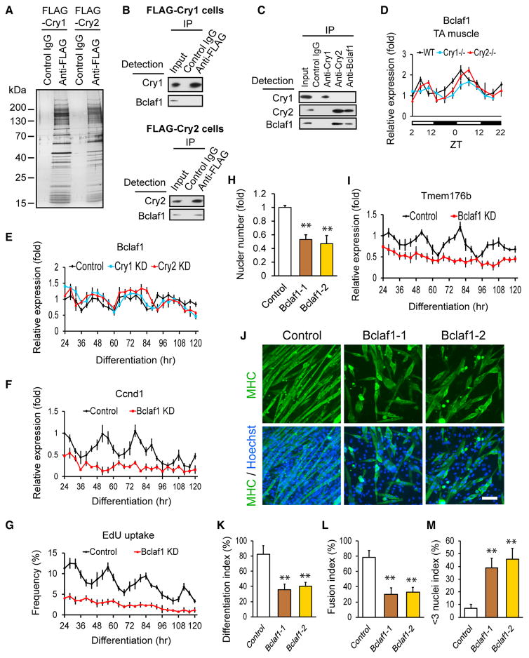
Figure 6. Regulation of Ccnd1 and Tmem176b by Bclaf1.
(A) Silver staining of a gel loaded with immunoprecipitated proteins from FLAG-Cry1- and FLAG-Cry2-expressing undifferentiated cells using an anti-FLAG antibody.
(B) Western blotting of immunoprecipitated proteins with an anti-FLAG antibody from FLAG-Cry1-expressing (top) and FLAG-Cry2-expressing (bottom) cells. Proteins were detected by the indicated antibodies.
(C) Western blotting of immunoprecipitated endogenous proteins with anti-Cry1, anti-Cry2, and anti-Bclaf1 antibodies from differentiation day 3 cells.
(D) Temporal profile of Bclaf1 expression levels in TA muscles.
(E–G) Temporal profiles of expression levels of Bclaf1 (E) and Ccnd1 (F) and uptake of EdU (G) in KD cells during differentiation. shRNA clone 1 was used for Cry1, Cry2, and Bclaf1 KD cells.
(H) Nuclear number in Bclaf1 KD cells on day 5.
(I) Temporal profiles of expression levels of Tmem176b in Bclaf1 KD cells during differentiation.
(J) MHC staining of Bclaf1 KD cells on day 5. Scale bar, 100 μm.
(K–M) Differentiation index (K), fusion index (L), and < 3 nuclei index (M) of Bclaf1 KD cells on day 5.
Data are presented as mean + or ± SD.

