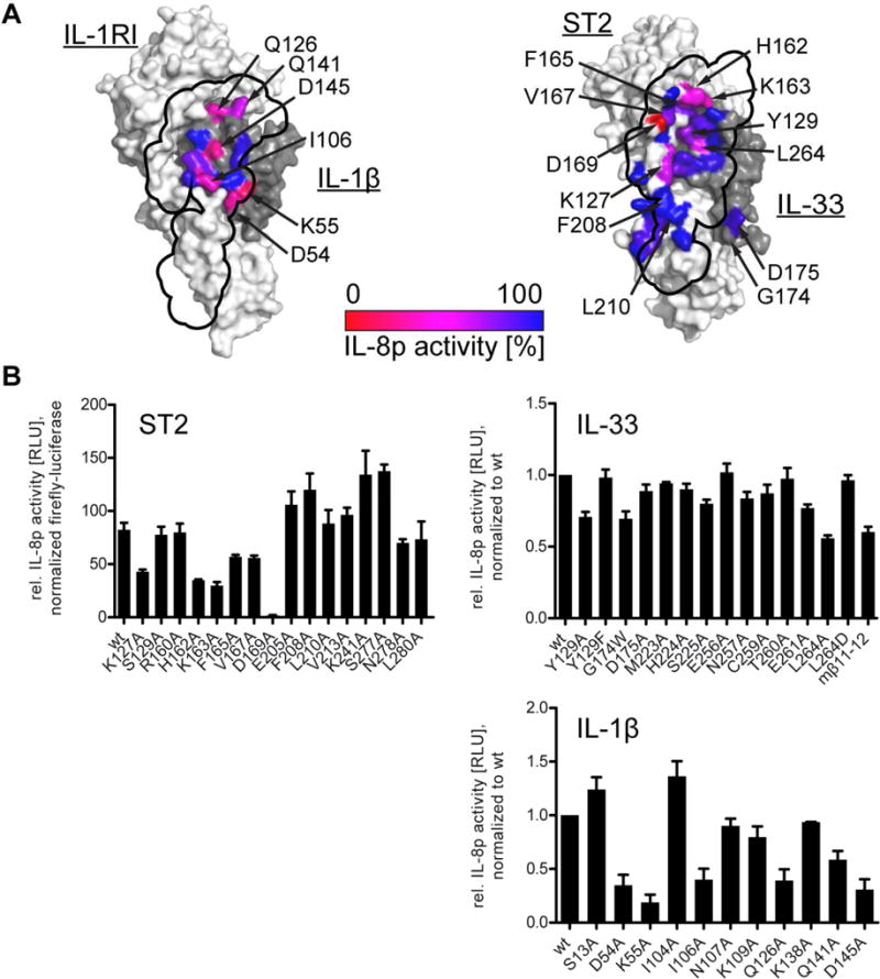
Fig. 7. IL-33 signaling depends less on direct cytokine:co-receptor interactions than IL-1β.

Signaling was assessed in HEK293T-based reporter cells containing an IL-8 promoter-driven luciferase. Cells were activated with either 5 pM IL-33 or 100 pM IL-1β and luciferase activity was measured 5 h after stimulation. A, location of mutants at the IL-1RAcP interface is indicated on the binary cytokine:receptor surfaces (left, IL-1β:IL-1RI; right, IL-33:ST2) and colored according to their activity relative to wild type signaling. The binding site of IL-1RAcP is projected onto the binary cytokine:receptor surface (black outline). B, results from signaling assays that form basis for coloring in A. For ST2 mutants (upper left), data were normalized to co-transfected, constitutively active firefly luciferase to normalization for transfection efficiency. For analysis of cytokine mutants (IL-33 upper right, IL-1β lower right), results were normalized to wild type cytokines. Data represent average of three independent experiments, each conducted in triplicate. Error bars represent SEM.
